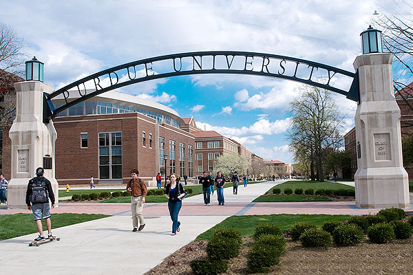
A Purdue University-affiliated startup, has devised a way to map arteries in the roof of a person’s mouth to help avoid complications and improve outcomes in oral surgery
Starfish Engineering has developed a method using light to image arteries and lesions through the tissue in the roof of the mouth. Surgeons working in the area of the mouth use the greater palatine artery as a landmark and need to know where it is to avoid damaging it or its surrounding nerves. But studies have shown that there can be a discrepancy of up to four millimeters between where surgeons believe the artery is and where it actually is, a disparity that can cause a variety of surgical complications and injuries.
A potential serious complication is hemorrhage. Significant bleeding can occur if the greater palatine artery is severed, especially if the artery retracts into the bony canal making it difficult to access. Most surgeons, therefore, justifiably avoid involving the entire range of possible locations of the greater palatine artery and surrounding area in the surgical field. However, due to significant individual variation, landmarks only provide a rough estimate of the actual location in a given patient. At present there is no available device to accurately identify the location of the foramen and artery in an individual patient in real time during surgery, and is therefore an unmet clinical need.
“This approach has the potential to reduce complications during surgery,” said Brian Bentz, Chief Executive Officer, Starfish. “It can help improve the accuracy of incisions.”
The process created by Bentz and Kevin Webb, a professor of electrical and computer engineering at Purdue, along with Vaibhav Gaind and Timothy C Wu, can be used in periodontal and oral surgeries, sinus and ridge augmentations, greater palatine nerve block, soft tissue biopsies, dental implants and wisdom teeth removal.
The process uses diffuse optical imaging to locate the greater palatine artery and lesions that are not visible to the naked eye by injecting light to create a 3-D map.
“The greater palatine artery is filled with blood so it will absorb more photons. You can use that contrast in absorption to determine the location of that artery,” Bentz said.
The specificity of lesions could be improved with topical fluorescence or absorption contrast agents, Bentz said.
The results of the complex shapes can be displayed on a mobile device or a computer. The information can be used to make printed surgical guides that can be placed on the roof of the mouth to help during surgery. The method also can be useful in identifying operative and postoperative bleeding.
- Advertisement -


Comments are closed.